Blood Flow Effects
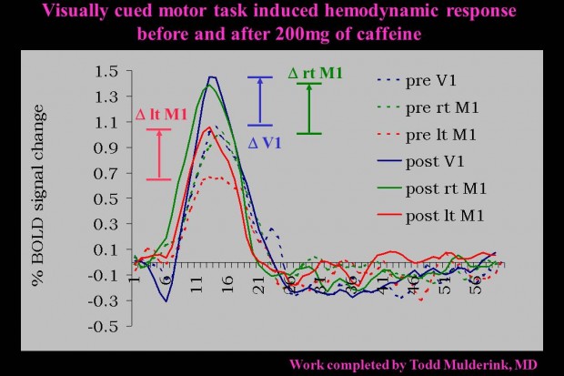
Some interesting work is underway investigating the effect of caffeine (a cerebral vasoconstrictor) on the brain, which may have the potential to be an fMRI contrast booster. The Parrish group was the first to explore the effects of caffeine on the BOLD signal in humans, see figure to the right. The early work demonstrated the dramatic gain (30-50% increase) in BOLD signal obtained by administering a dose of caffeine equivalent to just 2-3 cups of coffee. Read Mulderink Paper. However, caffeine is a vasoconstrictor which causes the resting perfusion level to decrease by ~20-30%. This led us to investigate some of the metabolic effects knowing that we perform better with caffeine but have reduced blood flow.

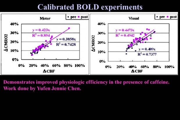
demonstration of the efficiency gained after taking caffeine
Metabolic Effects
Additional research conducted in the lab demonstrated that the brain physiology becomes more efficient at processing oxygen in the presence of caffeine, see figure to right. This figure demonstrates how the brain becomes more efficient in the presence of caffeine. That is the same level of metabolism (activation) post-caffeine (pink) requires a smaller blood flow change compared to the pre-caffeine condition (blue).
Effects on Resting State
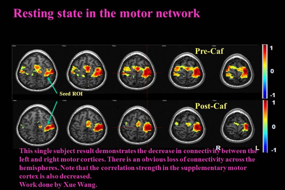
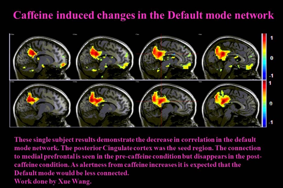
Current work is aimed at better understanding caffeine’s effect on the central nervous system with more complex tasks and additional information from other imaging modalities. Another topic of interest is how caffeine interacts with resting state connectivity measured with BOLD. This work demonstrates that there is dramatic change in connectivity measured by time course correlations of resting state data in different networks. These figures (see below) indicate that the BOLD resting state signal is a mixture of neuronal information complicated by physiologic noise, which is reduced in the caffeine condition. More work is required to better understand the meaning of resting state BOLD. (Read Caffeine CMRO2 Paper and Caffeine Dose Paper).
Future studies are aimed at investigating how caffeine can be used as a cognitive enhancer by facilitating learning or priming the cortex to receive therapy.

Caffeine’s influence on Resting State BOLD in the Motor network

Caffeine’s influence on the BOLD Resting State in the default mode network

