DTI Basics
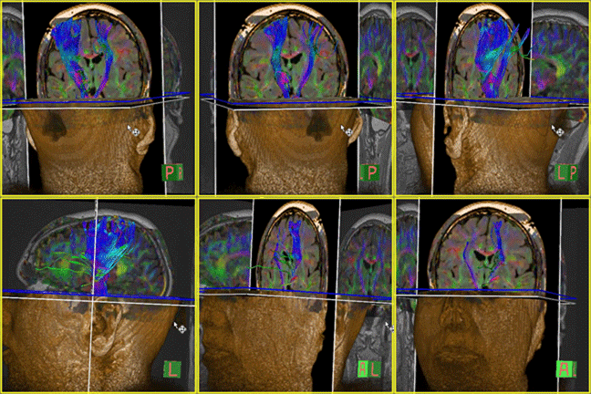
DTI exploits a property of MRI that makes the image sensitive to microscopic movement of water. As a result it is possible to explore white matter connectivity and physiologic properties. Beautiful images of localized white matter tracts are possible as well as being quantitative (See movie to the right).
We have utilized DTI metrics in the study of stroke subjects pre/post treatment to demonstrate the effect of aggressive treatment in chronic stroke. In addition, we are using DTI metrics to predict which stroke subjects would respond to therapy.
DTI for Parkinson’s Disease
A current study in collaboration with colleagues in the Department of Neurology (Drs. Simuni and Gitelman ) is investigating the use of DTI metrics as a biomarker for Parkinson’s Disease.
DTI of Learning
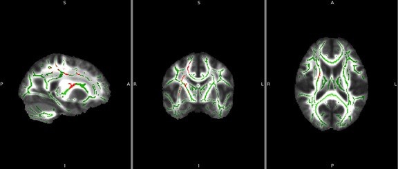
An exciting study that is in the early stages of development is the effect of learning on the cortical spinal tracts. A study with collaborators at the Rehabilitation Institute of Chicago (Mussa-Ivaldi, PhD) utilize a Brain Machine Interface to investigate the plasticity of the normal brain. Normal subjects were trained to use their shoulder muscles to guide a 3D cursor using an infrared interface to map the body position to a computer cursor movement. We used Tract based spatial statistics (TBSS) to calculate differences in white matter metrics compared to baseline. The results indicate an increase in the white matter coherence (FA value) in only the non-dominant corticospinal tract. (See red regions superimposed on the FA skeleton, below) This may be in response to a finer tuning of the non-dominant motor system.

The red regions indicate where the FA has increased following training. There were no places where the FA was larger at baseline compared to post-learning. TBSS skeleton in green.
These results demonstrate that it is possible to identify regions of the white matter that are involved in learning. Future work will investigate these same tasks in spinal cord injury patients.

