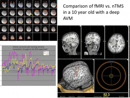
Results of the failed fMRI study in a 10 year old are shown on the left. On the right shows the successful results of the nTMS mapping, which showed traditional motor mapping requiring the surgeon to take a more conservative approach to treatment.
This relatively new method of non-invasively mapping brain function uses strong magnetic pulses to stimulate underlying cortex. The breakthrough in this technology has been the marriage of infrared neuronavigation with the TMS device (navigated TMS or nTMS). Now it is possible to know precisely where you are stimulating in the brain. Nexstim and the Parrish group have been collaborating to compare nTMS presurgical mapping to our clinical presurgical fMRI mapping using direct cortical stimulation intraoperatively as the gold standard. To date, the results of the study have shown that both methods match closely to the intraoperative findings. However, TMS is much less difficult to perform and does not require the patient to lie in a magnet. In fact, the case shown to the right demonstrates how we could not get a successful block motor fMRI study on a 10 year old AVM patient. The upper left of the figure shows a great deal of artifact in the functional map due to movement. The motion correction plot below the fmri maps shows how much movement is present in the experiment. However, the nTMS study was successful because he was able to watch Star Wars the Clone Wars, while being tested. Since TMS measures the tracts passively, the subject does not have to pay attention or even be awake (See figure to the right).

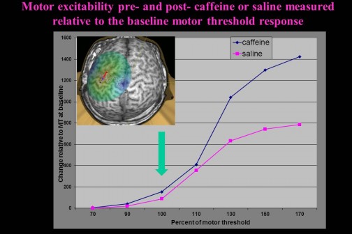
Transcranial Magnetic Stimulation (TMS) was used to map the motor excitability before and after caffeine. The curves indicate the EMG response following a specific stimulus intensity relative to the motor threshold (100%). Note how the post-caffeine condition (blue) is always larger than the placebo condition.
Another exciting study completed by high school students from the Illinois Math and Science Academy investigated the effect of caffeine on motor excitability. In their study, the students used navigated TMS to measure the response to a number of different stimulation levels pre and post caffeine. Due to the ability to precisely locate the magnetic stimulation pulse on the Nexstim system, it was possible to confidently stimulate at the same site. The results indicate that there is a >50% increase in the excitability of the motor neurons at the motor threshold level following caffeine and even a larger response at higher stimulation levels (still sub-threshold for overt movement). (See Figure to the right)
TMS measures purely neuronal stimulation compared to blood flow based effects in fMRI. Using these modalities together they give us a much better understanding of the complete effects of caffeine on the brain.
Future work will focus on mapping language areas for presurgical mapping and the investigation of therapy in aphasia subjects.
