Parrish Neuroimaging Group
  • Team
    • Todd Parrish, PhD
    • Jennie Yufen Chen, PhD
    • Xue Wang, PhD
    • Kenneth Weber, DC, PhD
    • Xiaowei Song
    • Ellen Fitzmorris, B.S.
    • Suvarna Badhe
    • Brian Williams, BSN, RN
  • Projects
    • Caffeine Research
    • Diffusion Tensor Imaging (DTI)
    • Temporal Signal to Noise in Time Series Data
    • Hemodynamic Response Function (HRF)
    • Transcranial Magnetic Stimulation (TMS)
    • Acupuncture
    • The Cerebrovascular Reserve (CVR)
    • Imaging of the Spinal Cord
    • Voxel Lesion Symptom Mapping (VLSM) in Chronic Stroke
  • fmri
    • fMRI Basics Presentation
    • Clinical
  • Volunteer
  • Contact Us
  • Neuroimaging Course
    • Neuroimaging Course
      • Registration
    • Advanced Neuroimaging: Fundamentals and Methods
Home» Research Projects » Voxel Lesion Symptom Mapping (VLSM) in Chronic Stroke

Voxel Lesion Symptom Mapping (VLSM) in Chronic Stroke

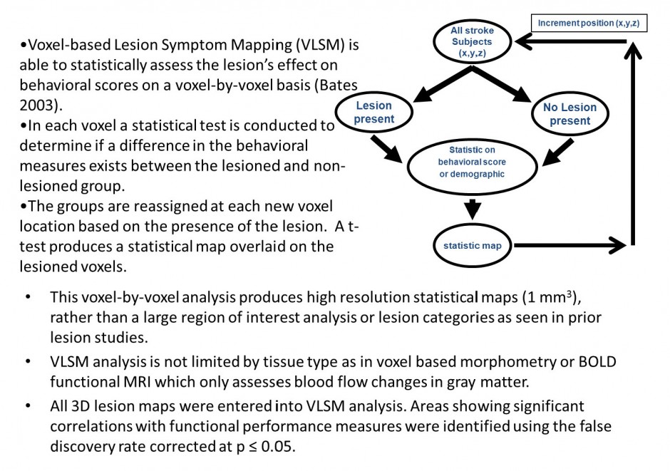
Voxel Lesion Symptom Mapping

  • Bates, E., Wilson, S.M., Saygin, A.P., Dick, F., Sereno, M.I., Knight, R.T., Dronkers, N.F. Voxel-based lesion-symptom mapping, Nat. Neurosci. 6, 448–450, 2003.
  • Lo R, Gitelman DR, Levy R, Hulvershorn J, Parrish TB. Identification of critical areas for motor function recovery in chronic stroke subjects using voxel-based lesion symptom mapping. Neuroimage 49(1), 9-18, 2010.

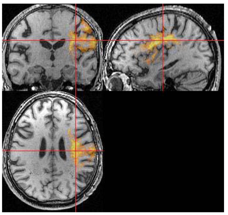
Voxel Lesion MRI

Data from all subjects (n=140) with lesions flipped into the left hemisphere. These data are FDR corrected p<0.05.

The area identified is the confluence of the sensory and motor signals that most likely integrate to produce the desired behavior. This region has been identified as a critical component of motor function following stroke. If a subject’s stroke involves these regions they are will have poor motor behavior.



(c) 2014 Parrish Neuroimaging Group