-
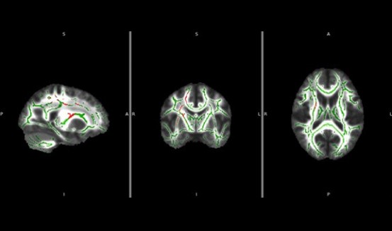
An exciting study that is in the early stages of development is the effect of learning on the cortical spinal tracts. A study with collaborators at the Rehabilitation Institute of Chicago (Dr. Musso-Invaldi) utilize a Brain Machine Interface to investigate the plasticity of the normal brain. Normal subjects were trained to use their shoulder muscles to guide a 3D cursor using an infrared interface to map the body position to a computer cursor movement.
-
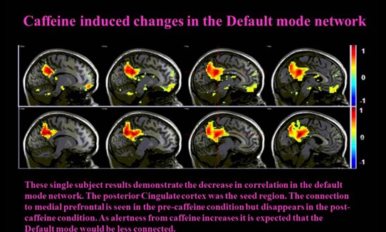
Some interesting work is underway investigating the effect of caffeine (a cerebral vasoconstrictor) on the brain, which may have the potential to be an fMRI contrast booster. The Parrish group was the first to explore the effects of caffeine on the BOLD signal in humans, see figure to the right. The early work demonstrated the dramatic gain (30-50% increase) in BOLD signal obtained by administering a dose of caffeine equivalent to just 2-3 cups of coffee.
-
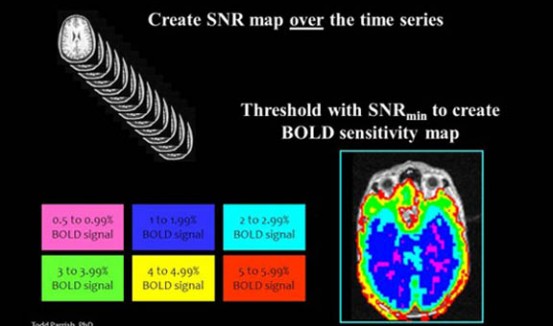
The signal detected in functional magnetic resonance imaging (fMRI) is on the order of a one percent from the baseline signal. Time series data with a high signal-to-noise ratio (SNR) is required to reliably detect these changes. This project developed a model to determine the minimum required signal-to-noise ratio necessary for detecting the expected signal change. The immediate benefits are clinical, addressing the neurosurgeon’s need to know if a region of brain near a lesion is active.
-
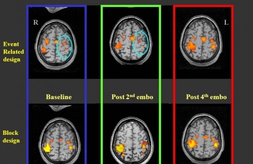
Another useful tool for investigating the underlying physiology is the impulse response function or the hemodynamic response function. Using brief visually cued motor stimuli, it is possible to measure properties of the local cerebrovasculature. This information is useful for normalizing blood oxygen level dependent (BOLD) signal or for detecting abnormalities.
-
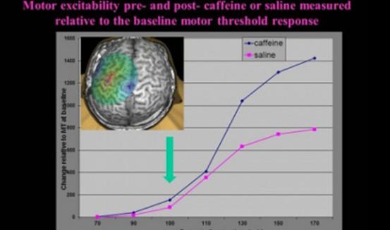
Another exciting study completed by high school students from the Illinois Math and Science Academy investigated the effect of caffeine on motor excitability. In their study, the students used navigated TMS to measure the response to a number of different stimulation levels pre and post caffeine. Due to the ability to precisely locate the magnetic stimulation pulse on the Nexstim system, it was possible to confidently stimulate at the same site.
-
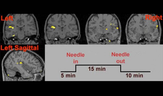
Acupuncture is a treatment for pain and illness in which thin needles are positioned just under the surface of the skin at special centers around the body. It originated in China over 3000 years ago, but despite its longevity Western medicine has been reluctant to accept acupuncture as a valid method of treatment. In our lab,we investigated the effect of acupuncture on the brain using functional magnetic resonance imaging (fMRI).
-
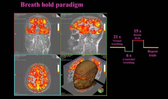
CVR is a quantitative measure of the cerebrovascular response to a challenge. Typically the challenge is breathing CO2 enriched air or being administered Acetazolamide, which causes an increase in the cerebral blood flow (CBF). We have implemented a method of breath holding to act as the vascular challenge.
-
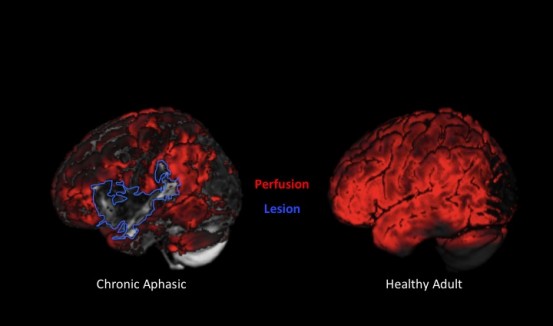
Individuals with the stroke-induced language disorder called aphasia can improve with language treatment, but the neurobiological mechanisms of recovery are unknown. In several subtypes of aphasia, capillary blood flow (‘perfusion’) in language regions of the brain has been linked with aphasic deficits and recovery. A new study investigates patterns of lesioning and perfusion, measured by arterial spin labeling (ASL) fMRI, and their impact on treatment-induced language recovery in chronic agrammatic aphasia.
-
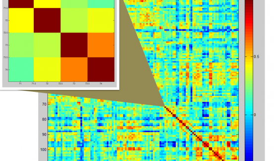
The connectivity matrix for 116 anatomic regions of interest was generated from resting state data from Parkinson’s disease patients. The enlarged image shows the connectivity matrix from a PD patient with low connectivity among the left Caudate, right Caudate, left Putamen, and right Putamen in the Basal Ganglia. The warmer colors show higher connectivity; whereas, the cooler colors show lower connectivity between brain regions of interest.
-
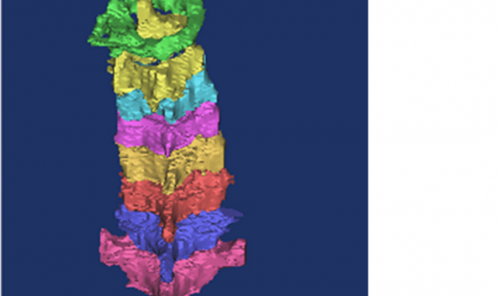
Data from a 3D MRI scan using a special positioning device was used to create 3D model of the cervical spine rotated 40 degrees to the left (view is from posterior). This work was done in collaboration with researchers in the Department of Orthopedic Surgery’s Biomechanics laboratory at Rush University. The aim of this work is to understand normal motion and how it changes in patients with neck pain.
-
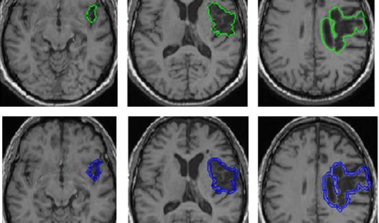
Brain lesions are typically hand-drawn slice by slice by trained professionals. It is a process that is time-consuming and prone to errors. Mean shift clustering is a method of image segmentation that can be used in conjunction with properties of brain symmetry to automatically delineate chronic stroke lesions. This is an example of a medium-sized brain lesion in the right hemisphere with a very high average Dice value (0.7869) compared to the hand-drawn gold standard.
-
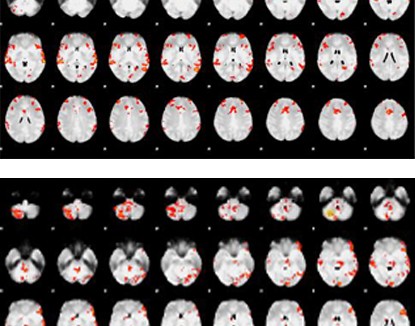
The top image shows activation of an individual with a normal brain who listened to a list of words and tones without any instructions. The bottom image shows brain activation from the same individual who listened to the same list of words and tones but pushed a button when he or she heard a pair of synonyms or the same tone. The similarities in activation of language processing centers support the hypothesis that individuals in a minimal conscious state may be capable of processing language. We can use these probes to investigate changes in patient’s level of consciousness.
Advanced Neuroimaging with a Clinical Focus
The primary focus of the Northwestern University Parrish Neuroimaging laboratory is the development and application of imaging methods to better understand human development and physiology.
Work in the lab draws from many disciplines such as biomedical engineering, biophysics, statistics, neuroscience, and physiology. While typical development occurs in normal healthy volunteers, a major effort is made to investigate the underlying physiology and function in a variety of disease conditions.
Message from the Director
Message from Todd Parrish, PhD
Contact Information
Contact Our Group
