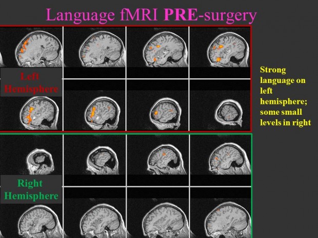
The use of functional magnetic resonance imaging (fMRI) for pre-surgical lesion mapping is an exciting field. Prior to fMRI, brain mapping has been accomplished during the actual surgical procedure and is limited to a hemisphere. Furthermore,using fMRI it is possible to assess multiple “tasks” spanning different domains of function.
Before fMRI, the most important information – assessment of the contra-lesional hemisphere of the brain – was unavailable. The contra-lesional hemisphere was never exposed during surgery and thus was not tested. With fMRI, the entire brain can be studied.
Since 1996, we have studied several hundred patients who have had tumors or arteriovenous malformations (AVM). An AVM is caused when high-pressure arterial blood is shunted directly into the low-pressure venous system. AVM patients are at high risk for a vascular rupture. The abnormal blood flow also has an impact on the local brain function (See HRF project page). Since these lesions are believed to be congenital, the brain has the ability to “rewire” the circuitry to account for the presence of the lesion.
We have found that in a number of patients with AVMs the contra-lesional hemisphere is involved in the performance of a task that should be controlled by the hemisphere containing the AVM. This can potentially have a positive impact on the surgical procedure. In fact, it could make it possible for somebody who is not eligible for treatment to actually obtain it.
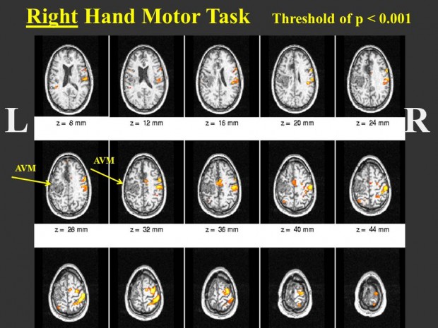
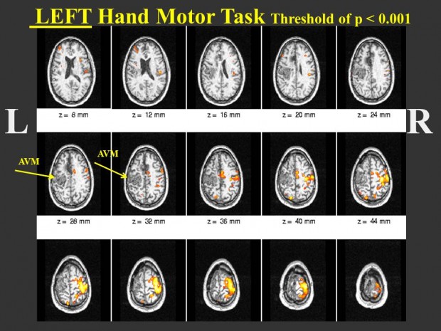
For example, (see Figure 1) when a patient with an AVM in the left hemisphere performs a left hand motor task, focal activation in the motor and sensory areas is seen in the right hemisphere, indicating a consolidated use of the cortex. When the same patient performs a right hand motor task (which should activate the left motor cortex), no activation in the left hemisphere is observed (Figure 2). However, there is strong activation in the right hemisphere in regions of pre-motor and sensory cortex. The presence of the AVM may be disturbing the flow making it difficult to detect activation around the AVM, and the right hemisphere activation may represent cortical re-mapping.
Pre and Post Tumor Surgery
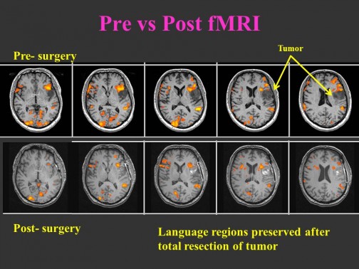
Pre- and post-surgery in a tumor patient is shown in Figure 3. Notice how the language activation is preserved. In the post-surgical imaging there are blood by-products present in the resection cavity which show increased signal due to the reduced T1. This patient went home 2 days after their surgery with their language intact.
Pre and Post AVM Surgery
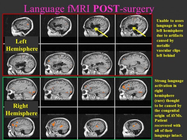
Figure 4 depicts the left and right hemisphere in a patient with a medium-sized AVM in the left temporal lobe. The pre-surgery fMRI study demonstrates strong left lateralized language with some weak activation in the right hemisphere. Figure 5 shows the post-surgery fMRI map. Note that there is serve artifact in the left hemisphere due to the presence of metallic clips used to treat the AVM. Therefore, we cannot assess the status of language in the left hemisphere. However, it is clear that language activation is present in the right hemisphere. It could be the congenital nature of the lesion made it possible to transfer language function to the right hemisphere.

